Currently the group hosts
a SPECT prototype and a PET prototype unique on national and regional
level.
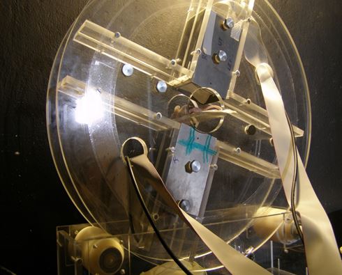
The SPECT system was constructed by the group of Dr. Stan Majewski in Jefferson Lab (US), in terms of a common cooperation. The system is based on 2 PSPMTs a pixilated NaI(Tl) scintillator, a parallel hexagonal hole collimator and a computer controlled gantry. The system has a 10x5cm Field of View (FOV). Its spatial resoliution is 1.5mm in scintigraphic mode and ~2mm in tomographic mode. The energy resolution has been measured equal to ~15%. This system has been used in numerous small animal studies over the past five years.
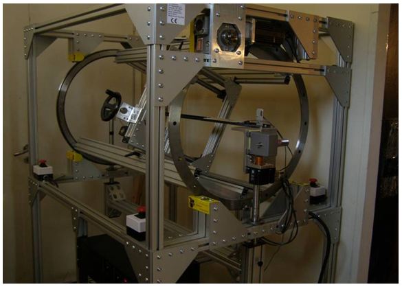
The PET system was a result of the same collaboration; It has a field of view of 5x5cm and is based on 2 H8500 Position Sensitive Photomultipier Tubes (PSPMTs), coupled to two LSO crystals with 2.5x2.5mm pixel size. PET data are obtained by rotating the two heads using a custom made gantry. The system has been used in the first PET animal studies in Greece.
Both systems are installed at the Institute of Nuclear and Radiological Sciences and Technology, Energy & Safety at N.C.S.R. ‘Demokritos Research Center' in Athens, Greece. In terms of a National Excellence project both systems are now being upgraded. The existing 2-head PET prototype will be redesigned and the field of view will be extended to 5x10cm. In addition an X-ray system will be purchased. The two detectors will be combined with the existing SPECT system and will result to a unique SPECT/PET/CT protoype.
The SPECT system was constructed by the group of Dr. Stan Majewski in Jefferson Lab (US), in terms of a common cooperation. The system is based on 2 PSPMTs a pixilated NaI(Tl) scintillator, a parallel hexagonal hole collimator and a computer controlled gantry. The system has a 10x5cm Field of View (FOV). Its spatial resoliution is 1.5mm in scintigraphic mode and ~2mm in tomographic mode. The energy resolution has been measured equal to ~15%. This system has been used in numerous small animal studies over the past five years.
The PET system was a result of the same collaboration; It has a field of view of 5x5cm and is based on 2 H8500 Position Sensitive Photomultipier Tubes (PSPMTs), coupled to two LSO crystals with 2.5x2.5mm pixel size. PET data are obtained by rotating the two heads using a custom made gantry. The system has been used in the first PET animal studies in Greece.
Both systems are installed at the Institute of Nuclear and Radiological Sciences and Technology, Energy & Safety at N.C.S.R. ‘Demokritos Research Center' in Athens, Greece. In terms of a National Excellence project both systems are now being upgraded. The existing 2-head PET prototype will be redesigned and the field of view will be extended to 5x10cm. In addition an X-ray system will be purchased. The two detectors will be combined with the existing SPECT system and will result to a unique SPECT/PET/CT protoype.
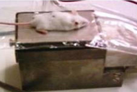
| Head of the SPECT system and planar mouse imaging | Existing 5x10cm SPECT system |
Existing 5x5cm PET system |
 |
 |
 |
Static bone image of a
mouse using 99mTc-MDP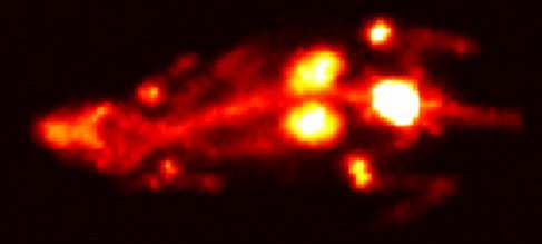 |
Tomographic imaging of a
mouse injected with 99mTc-labelled nanoparticles |
Successive slices of
a mouse injected with FDG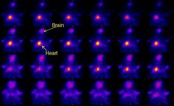 |
.