We have designed and constructed a tomographic imaging system, suitable for small animal SPECT imaging, which is optimized for rat and rabbit imaging. The system has been installed in 2013 at the Medical Shool of University of Patras.
The camera head consists of: 4 H8500 PSPMTs, a CsI:Na with 1.5x1.5x6mm3 pixel size, a parallel hole collimator 26mm in height, 1.2mm hole with 0.16mm septa. The readout electronics for reducing the 256 input to 4 output signals and amplyfing them are designed and made by our group. The data acquisition system is based on free-running sampling ADCs, FPGA electronics and an ethernet protocol for data transmission to a standard laptop. The gantry has a diameter of rotation equal to 15cm (for rabbit and rat imaging). A stepper motor is controlled by an AVR microcontroller via USB communication. Custom software was developed for motor rotation control, data acquisition and tomographic reconstruction (QSPECT open-source software, developed by our group).
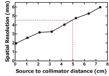
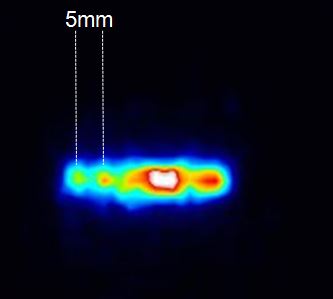
The spatilal resolution of the system is 1.9mm on the collimators surface and 5mm at 5cm distance in planar and tomographic mode. The sensitivity of the system was measured equal to ~110cps/MBq using a capillary source. The energy resolution was measured equal to 34% using a 99mTc flood source in front of the collimator.
|
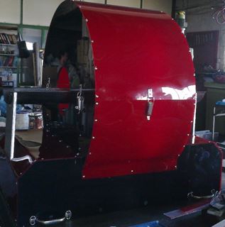
Full SPECT system
|
Systems spatial
resolution
|
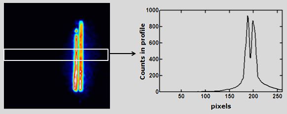
2 capillaries filled with 99mTc at 3.4mm distance |
 |
 |
 |
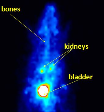
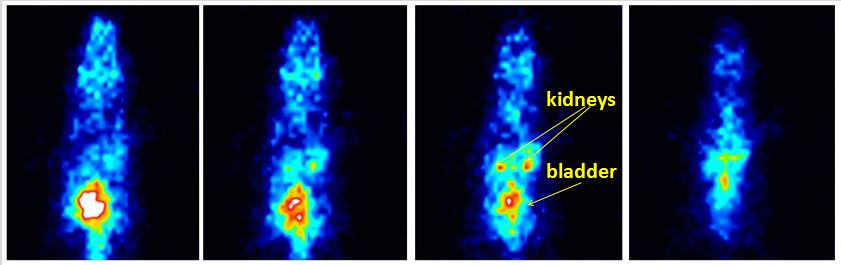
| Scintigraphic image of a mouse injected with 99mTc- DMSA | Tomographic slices of a capillaries filled with 99mTc | Tomographic slices of a mouse injected with 99mTc- DMSA (camera rotates at 7.5cm distance) |
 |
 |
 |
.